
Skull Base Tumors
Successful surgery demands the use of advanced, high-quality operating tools such as operating microscopes, smooth and safe anesthesia, intraoperative neuro-monitoring, facilities for postoperative ventilation, and a highly experienced multi-disciplinary surgical team. These tumors develop typically in deep and sensitive locations near critical blood vessels and nerves, making the procedure complex and demanding.
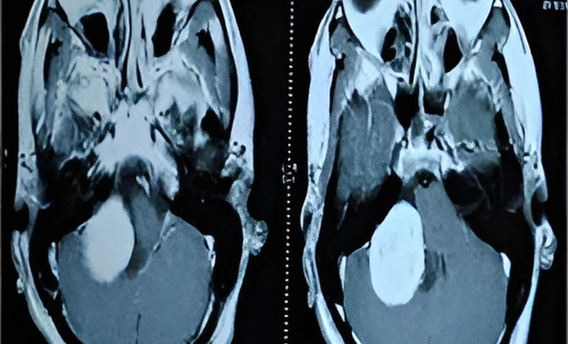
Symptoms and Diagnosis
Symptoms depend on tumor size, location, and nerves affected, including headaches, facial weakness or numbness, vision changes, hearing loss, difficulty swallowing or speaking, balance problems, and hydrocephalus-related symptoms like nausea and vomiting. Diagnosis usually involves detailed neurological examinations along with imaging modalities like MRI and CT scans to map tumor extent and involvement of adjacent structures.
Frequently asked questions
Tumors that grow in the skull base near vital nerves and blood vessels, often large and complex.
Symptoms vary but commonly include headaches, cranial nerve deficits, vision and hearing problems, and hydrocephalus.
Because tumors lie deep near delicate neurovascular structures, and surgery requires precision, advanced tools, and experienced teams.
Operating microscope, neuro-monitoring, smooth anesthesia, specialized operating tables, and post-op ventilation support.
Through neurological exams and detailed imaging like MRI and CT scans.