
Malignant Glioma
Diagnosis relies on advanced imaging techniques such as MRI and CT scans to visualize the tumor and its effects within the brain. Biopsy and molecular testing provide a definitive diagnosis and guide treatment decisions. Common symptoms include seizures, personality changes, loss of coordination, persistent headache—most noticeable in the morning—vision problems, and speech difficulties.
Treatment Approach and Results
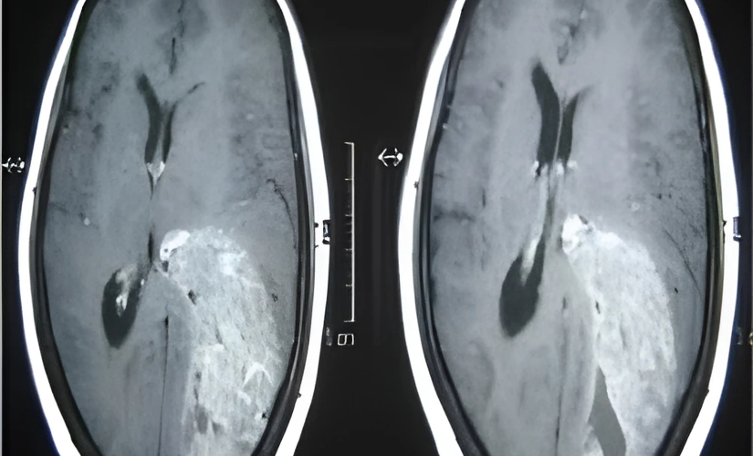
Treatment typically begins with surgical excision of the tumor, aiming for maximal removal while minimizing damage to healthy brain tissue. Advanced techniques, such as fluorescence-guided surgery, enable more precise excision. Post-surgical imaging (CT/MRI) confirms the removal. Patients then receive radiotherapy and chemotherapy to target any remaining cancer cells. Follow-up imaging helps monitor recurrence, and a clear MRI one year post-treatment, as in this referenced patient case, indicates effective management and an encouraging prognosis
Frequently asked questions
Headaches—especially in the morning—neurological deficits, seizures, personality or memory changes, visual problems, and speech impairment
Using CT/MRI imaging followed by biopsy and molecular analysis to confirm tumor type and grade.
A technique in which special dyes highlight tumor cells under specific lighting during surgery, enabling surgeons to excise the tumor more completely.
Yes. These are vital for destroying residual cancer cells and reducing the risk of recurrence, and are tailored to each patient.
While prognosis varies by tumor type, grade, and treatment response, aggressive surgery and adjuvant therapy improve survival and quality of life.